
Nail Anatomy
Authors: Doug Schoon and Ana Seidel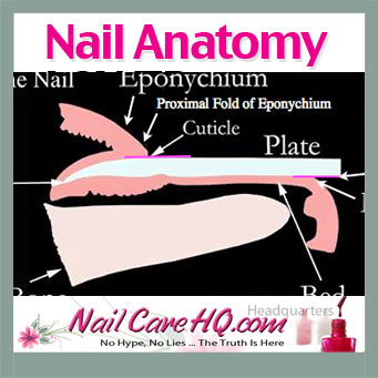
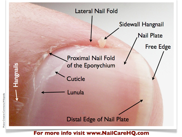
Nail Anatomy – The Different Parts of the Fingernail
Do you know where your cuticle is? Or your hyponychium? Most people don’t.
Not only is the general public confused about the names for the parts of the natural nail, but many nail technicians are not able to name the various major parts and know their function.
Let’s change this today.
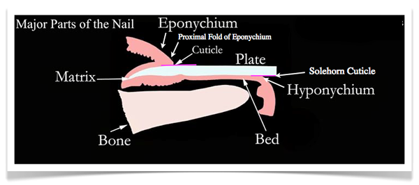
Matrix
Where new nail plate cells are created and the nail plate begins to form.
Lunula
Lunula
A bluish-white, opaque area that is visible through the nail plate. This area is the front part of the nail matrix. Sometimes, it’s called the “moon.”
The lunula is the front part of the matrix we can see, or in other words, the visible matrix.
Not all fingers have a visible lunula. Usually, it is easiest to find a lunula on a thumb or index finger.
Many people think that they would like to have lunula’s, but in fact, you really don’t.
Since it is the exposed portion of the matrix, this area is not protected by the eponychium. It is easy bruised with every day life tasks.
Those bruises show up as little white marks in the nail plate.
Eponychium
Living skin at the base of the nail plate that covers the matrix area. This should NOT be confused with the “cuticle”.
Proximal Fold of the Eponychium
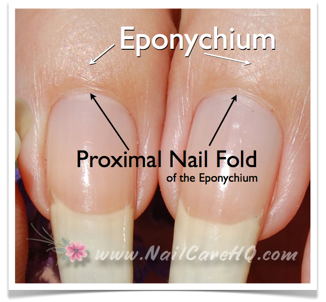
Healthy Proximal Fold
A tight band of living tissue that most people incorrectly think is their “cuticle”.
Since this skin dries out easily, people are quick to clip this skin believing that it’s not necessary.
Please, please, please…DO NOT CUT THIS SKIN!
The proximal fold is a required guardian seal that prevents germs and bacteria from getting to the nail matrix, where new cells are created.
I always know when girls are cutting. Their entire cuticle line is red and inflamed. Basically, their eponychium is infected all the time.
If you go to a salon for a manicure, do not ever let your nail tech cut this skin.
The best way to keep this skin soft and tight to the nail plate is with a high quality, jojoba wax ester based penetrating nail and cuticle oil.
Cuticle
*Represented as a pink line in the first photo
CLICK PICTURE FOR LARGER DETAIL
A thin layer of dead tissue riding on the nail plate to form a seal between the nail plate and eponychium to prevent pathogens from infecting the matrix area.
The cuticle pulls away from the underside of the eponychium and attaches tenaciously to the nail plate.
The cuticle should NOT be confused with the “eponychium”.
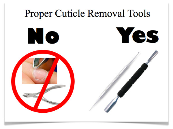
Since polish and nail enhancements don’t bond to skin on the nail plate, the cuticle should be properly removed with gentle scraping only.
Nippers can’t remove the cuticle. It’s physically impossible.
Nail Plate
Composed of hardened, flat, translucent, non-living, keratin nail cells that form a solid, protective layer over the underlying soft tissue.
The nail plate should NOT be confused with the nail “bed”.
The average person has 50 layers of keratin cells that make up the nail plate.
The thickness of your nails is determined by the size of your matrix.
Not everyone’s matrix is the same size. People with thin nails have a small matrix and will have less than 50 layers. People with thick nails have a large matrix and have more than 50 layers.
Nail Bed
The soft, pink tissue that sits underneath and supports the nail plate while it grows. The nail bed should NOT be confused with the nail “plate”.
CLICK FOR MORE DETAIL
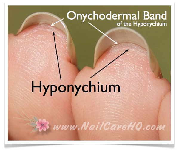
Hyponychium
*Represented as a pink line in the first photo
A soft tissue seal underneath the extended “free” edge of the nail plate whose purpose is to prevent pathogens from infecting the nail bed.
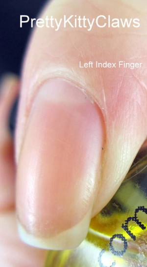
Onychodermal Band
A band of bunched up tissue located behind the hyponychium.
This band improves the ability of the hyponychium to prevent pathogens from infecting the nail bed.
The onychodermal band works in the same way as the proximal fold on the top surface of the nail.
When looking at your bare nails, you can see this darker band of skin right before your nail plate leaves the nail bed to become your free edge.
Solehorn Cuticle
A thin layer of dead tissue riding on the nail plate to form a seal between the nail plate and hyponychium to prevent pathogens from infecting the nail bed.
The solehorn cuticle pulls away from the underside of the hyponychium and attaches tenaciously to the nail plate.
Bone
Bone supports and shapes both the nail matrix and nail bed. The flat or curved shape of your nails is determined by the shape of your fingertip bone.
Knowledge is Power
Knowing your nail anatomy is important for the home manicurist and can actually help you find an excellent nail professional if you’re wanting to be pampered.
Ask her how she removes the cuticle.
If she shows you nippers instead of a spoon shaped remover or curette, you’ll know to politely walk out and find someone new.
I found this to be very educational, Anna and it directed me to find a new nail salon, where nippers to my proximal fold is amongst the first thing they want to do.
Professionals that don’t use correct names are insulting to their customers..They assume we aren’t intelligent enough or don’t care enough to know about our own bodies.
I did walk out of my local salon-where they can’t keep a tech more that a couple of months for some reason and when the owner asked why-I told him.
It may be close but I want a tech who respects my desires.
Thabks so much
Elle
This article was very helpful. I wish i read this earlier. I often end up cutting my proximal nail fold while cleaning up dead white tissues / cuticles. As mentioned in the article, yes, my nails remain red and in pain for couple of days till the skin is healed. Any recommandation which cuticle remover works without need of cutting free edge skin?
I use Sally Hansen cuticle remover about every two weeks. Make sure it does not touch your skin as much as possible, and you only apply it to the nail plate. You can use an orange wood stick to very, very gently scrape the skin off your nail plate. Do one hand at a time, and be sure to rinse it off as soon as you are done. Then move onto the second hand.
This article was ALL I needed to convince me I no longer want to go to the nail salon. I’ve been a home manicurist for a while but NOW I know why and how to properly care for my nails… I do better than the salon anyway thanks to you and your amazing website! THANK YOU ANA!
I feel so informed now! I’ll be sure to put manicurists in check if I see them with nippers trying to cut cuticles!
Hi, Ana
I have never really worried about my hands and nails until I had chemotherapy for breast cancer last year. The chemo damaged my nails really badly, and they went really brown and became very brittle, flakey and peeling. Some of them even started lifting off the nail bed. I couldn’t afford to buy your cuticle oil because of the postage costs (I was broke after all the cancer treatment), but I did get some jojoba oil, and have been using this, along with other cuticle oils and nourishing treatments. The new growth on my nails is looking OK, but the brittle part of them is still the same and hasn’t completely grown out. Also, the nails have still not attached back onto the nail bed. Could you tell me if my nails will ever return to the way they were before my treatment? Have you had any feedback from people like me who have used your cuticle oil? We were given no advice before having chemo on how to look after our nails – just told that we should paint them with dark nail polish to keep the sun off them and to cover up any damage that was done. If I had known how to properly care for my nails, and the proper way to do a manicure, before my treatment, I’m sure the damage would have been nowhere near as bad as it has been. Each time I do my nails I file a little more of the dark, horrible fragile end off, and at the moment I have one nail that broke 2 days after I did my last manicure, and one nail held together with the “tea bag” method, which I have been nurturing for about a month now. It is so fragile that, if I didn’t put the tea bag patch on it across the tip, it would have broken right off at the hyponychium, and of course that would hurt, and could open it up to infection. I would appreciate any advice you can give me that may help to improve my chances of getting my nails back to normal, or even better if I can, because they’ve never been very good. I bit them for over 30 years, which of course didn’t help them at all. Once I’ve recovered from the cost of Christmas, I hope to be able to buy some of your Pure Oil, but I wish it was available in Australia somewhere. If you would like me to test and review it for you, I would be quite willing to put it on my blog, which I have started to try and help others to stop getting the severed damage I did from the chemotherapy. It has become a crusade for me, and I’m even trying to get nail care advice added to the pre-chemo advice given to cancer patients. Thanks for all the wonderful advice you have given us, and for taking the time to share it with us.
Any health issues and treatments can be brutal to nail health.
Chemo is about killing cancer cells, so it’s going to kill a lot of other cells as well. Your nail matrix cannot possibly create good-quality cells. It takes nails four months to replace themselves, so you usually experience the results well after the treatments.
Since I’m not a doctor, I can’t make any diagnosis. But my assumption would be that once your health has improved, you will see that 4 months later.
Now I know nails are for more than picking up things. Ana, your advice is priceless, everyone should be informed on this very important part of the anatomy.
Respectfully Yours D.Ed.
Thank You! For this article, it is so educational and helpful.